


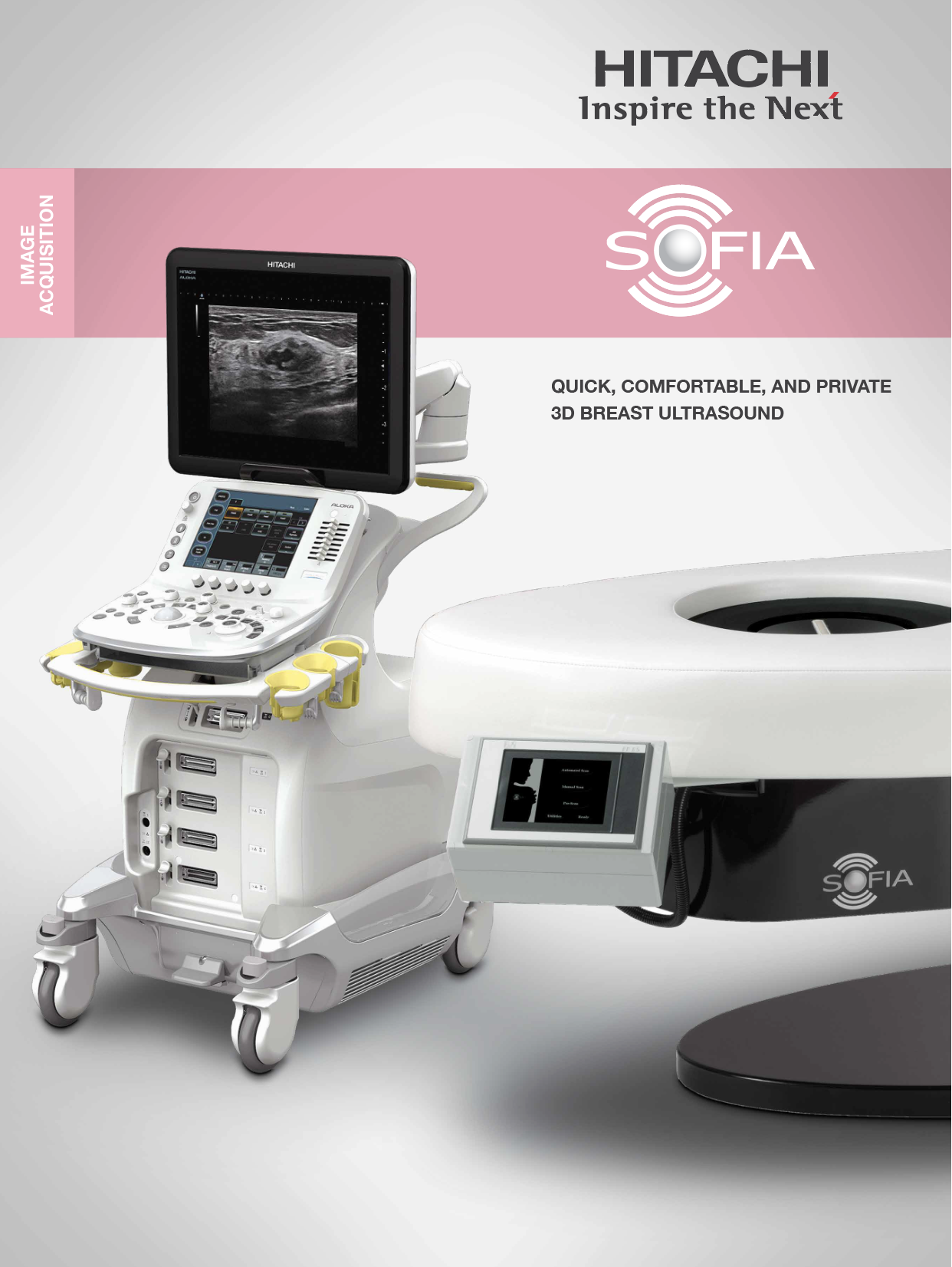
Efficiency and speed define SOFIA’s image interpretation process. Each breast is presented to the radiologist as a single, high-resolution DICOM-compatible dataset, interpreted in familiar planes. This streamlined approach significantly reduces review times.
Bella’s Battle is proud to introduce SOFIA, bringing precision, efficiency, and comfort to breast cancer diagnostics, setting new standards in the field.

A comprehensive axial view revealing a spiculated shadowing mass, confirmed as invasive ductal carcinoma through biopsy.
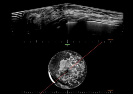
Top Image: A radial view of the left breast, prominently showcasing a lobule and its ducts extending outward from the nipple.
Bottom Image: The coronal view, positioned strategically to pinpoint the exact location of the radial slice.

A full-field radial image capturing the left breast’s condition, displaying multiple substantial cysts. This image, acquired from around the 9:00 to 3:00 positions of the breast, reveals a prominent cyst, accompanied by a solid mass positioned directly in front of it.
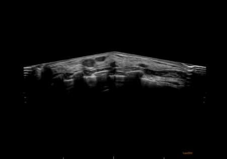
Complete view of a breast displaying multiple cysts and benign fibroadenomas. The signal reduction on both edges of the image corresponds to the lateral margin and the sternum.

An image showcasing a fibroadenoma viewed from three different angles simultaneously. The breast’s volume rendering (top-left) has been adjusted to show the patient’s ribs facing the viewer.